


Best Melasma/ Hyper-Pigmentation Treatment in Delhi at DermaWorld
Get the world-class Melasma or Hyper-pigmentation treatment in Delhi. DermaWorld, Delhi, headed by specialist Dermatologist Dr. Rohit Batra who does not profess but has been docketed providing specialized tailor-made procedures for Melasma/Hyper-pigmentation with above 90% of success rate as compared to those who are just indulged in money-spinning making big claims & delivering as low as 20%-30% success rate.
Why Superficial Hyper-Pigmentation Blasting Backfires
- Using high-energy lasers blindly on active, vascularized dermal melasma patches.
- Relying on generic over-the-counter bleaching creams that trigger ochronosis.
- Applying deep chemical peels without stabilizing baseline melanocyte activity.
- Treating systemic pigment issues as surface-level cosmetic abnormalities.
Dermal hyper-pigmentation is a complex cellular defense mechanism, not a simple surface stain. Securing the best Pigmentation Treatment in Delhi demands a highly structured, multi-tier intervention that systematically targets the tyrosinase enzyme while completely preserving epidermal integrity.
The Cellular Pathophysiology of Cutaneous Pigmentation
Melanogenesis and Tyrosinase Enzyme Activation
Cutaneous hyperpigmentation is a multi-factorial biological response driven by specialized dendritic cells located in the basal layer of the epidermis, known as melanocytes. These cells produce melanin, a complex bio-polymeric pigment synthesized inside specialized cellular organelles called melanosomes.
This process, known as melanogenesis, is controlled by an enzyme named **tyrosinase**, which oxidizes the amino acid L-tyrosine into DOPA and subsequently into dopaquinone.
When your skin encounters external stressors, it over-stimulates this cellular pathway. This causes an overproduction of pigment that is distributed to neighboring surface skin cells, leading to localized patches of dark skin.
Differentiating Superficial Staining from Deep Dermal Deposition
In clinical dermatology, treatments must distinguish between different types of pigment deposition. Superficial abnormalities, like sun-induced ephelides (freckles) and solar lentigines (age spots), feature localized pigment accumulation wrapped securely inside the upper epidermal layers.
In contrast, generalized post-inflammatory hyperpigmentation (PIH) and multi-layered patches like melasma involve a deeper, vascularized component hidden within the reticular dermis. Treating these deep structural conditions requires specialized, clinical-grade systems. Superficial beauty treatments cannot safely reach or clear these deep pigment deposits.
Systemic Melasma Treatment: Navigating Cellular Triggers
Hormonal Receptors and Environmental Stressors
Melasma is a complex, chronic condition characterized by symmetrical, dark brown patches that typically appear on the cheeks, forehead, upper lip, and nose. Unlike temporary sun damage, a professional Melasma Treatment in Delhi must address a delicate combination of hormonal, vascular, and environmental triggers.
The condition is highly sensitive to estrogen and progesterone levels, which directly up-regulate melanocortin receptors on the surface of pigment-producing cells. This hormonal baseline makes the cells hypersensitive to environmental factors like ultraviolet (UV) rays and high heat.
Furthermore, local atmospheric factors—such as seasonal high temperatures and intense levels of urban air pollution—generate substantial amounts of free radicals that damage cell membranes through lipid peroxidation.
This continuous environmental stress triggers a chronic inflammatory response within the upper skin layers, releasing chemical signals like prostaglandins and nitric oxide.
These inflammatory molecules stimulate nearby blood vessels, causing a condition known as **dermal angiogenesis** (the growth of micro-capillaries). These tiny blood vessels feed nutrients directly to the overlying pigment cells, creating a loop of pigment production. Therefore, trying to clear melasma with aggressive surface scrubs or high-heat lasers can irritate these blood vessels, causing a severe rebound of hyperpigmentation.
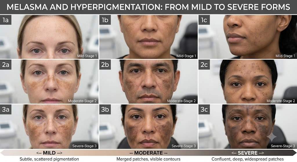
Mapping Melasma Across Severity Spectrums
Classifying pigmentation requires assessing whether the melanin deposition is confined to the superficial epidermal layer or extends deep into the reticular dermis with accompanying vascular expansion. Mild presentations respond readily to barrier stabilization and mild chemical pathways, whereas severe chloasma requires advanced energy devices.
By identifying your specific severity tier under the guidance of our trained Dermatologist, we prevent aggressive thermal damage and align our interventions with specialized resurfacing modalities like our Cosmelan Peels portfolio.
| Clinical Technology | Targeted Condition | Primary Operating Mechanism | Fitzpatrick Safety Strategy |
|---|---|---|---|
| Picosecond Q-Switched Nd:YAG | Deep Dermal Melasma & Post-Inflammatory Over-pigmentation | Photo-Acoustic Fragmentation | Delivers zero thermal effect to break up ink-like pigment into micro-dust without activating melanocytes. |
| Fractional Thulium Laser (1927 nm) | Resistant Epidermal Pigment & Sun Damage | Sub-Ablative Dermal Resurfacing | Creates microscopic treatment zones while fully leaving the stratum corneum intact to speed barrier healing. |
| Modified Kligman’s Chemical System | Refractory Chloasma & Superficial Staining | Synergistic Tyrosinase Inhibition | Combines hydroquinone with topical tretinoin to accelerate cell turnover while suppressing fresh pigment generation. |
| Tranexamic Micro-Injections | Highly Vascularized Melasma Overgrowth | Plasminogen Blockade | Suppresses specific chemical signals to shrink underlying blood vessels feeding the pigment cells. |
Managing Pigmentation Variations Across the Indian Continuum
Phototypes IV to VI and the Reactivity Index
A common medical error in non-specialist aesthetic clinics is treating all dark marks with a single, aggressive treatment format. Within the framework of medical dermatology, treatment settings are strictly determined by your specific skin profile on the Fitzpatrick Phototype Scale.
The majority of individuals across North India feature skin types falling between categories IV and VI. These darker, melanin-rich profiles feature highly active pigment cells that respond quickly to thermal shifts, physical irritation, or aggressive chemical applications.
If a clinic applies a high-energy laser setting or an aggressive chemical acid to a Fitzpatrick Type V profile, the underlying pigment cells identify that action as a severe threat. This triggers a defensive response known as rebound post-inflammatory hyperpigmentation, causing a worsening of dark patches across the face.
Comprehensive Dermal Care Protocols and UV Shielding
Post-Treatment Recovery and Barrier Maintenance
The permanent resolution of complex skin darkening requires a coordinated balance between advanced, in-clinic treatments and strict home care. Following an advanced session, such as a customized peeling treatment or a precision laser pass, the skin enters a sensitive recovery window.
During this initial phase, the outer layer is exceptionally vulnerable to ultraviolet radiation, which can quickly restart the pigment production pathway. To protect this fresh, healing tissue, patients must adhere to a detailed home skin care regimen.
A proper maintenance routine centers around the daily use of broad-spectrum physical sunscreens formulated with medical-grade zinc oxide or titanium dioxide shields. These minerals form a physical barrier that reflects harmful UVA and UVB rays away from the skin surface.
Additionally, incorporating morning antioxidant serums—like stable silymarin or ferulic acid—neutralizes pollution-induced free radicals, while evening applications of non-irritating brighteners like azelaic acid or kojic dipalmitate safely maintain low enzyme activity. Consistently following these steps ensures long-term skin clarity, helping you get the absolute most out of your advanced Medi Facial treatment in Delhi.
Dermal Melanogenesis Down-Regulation
Permanent pigment clearance operates deep within your cellular architecture. By choosing targeted, photo-acoustic fragmentation over high-heat lasers, we break apart trapped pigment deposits while protecting your natural skin tones.
Every step of our protocol is carefully calibrated to slow down overactive pigment production channels, protecting vulnerable surface layers from thermal stress or rebound darkening.
Dermatologist-prescribed active complexes block tyrosinase pathways to stop fresh pigment synthesis.
Picosecond acoustic pressure waves break up deep, dense melanin clusters into ultra-fine particles without generating heat.
Your body’s natural lymphatic system clears away the microscopic pigment dust, leading to progressive skin clearing.
Ready for Clear, Spotless Skin?
Entrust your pigmentation and melasma clearance journey to board-certified medical specialists. Stabilize your melanocyte activity safely without thermal rebound risks.